Home »
SMILE Pro Laser Eye Surgery
Written and edited by the Smart Laser Eye Center Editorial Staff · Medically reviewed by Dr. Alim Huseynov, MD · Last updated: 10 September 2026
SMILE Pro is a flapless laser vision correction procedure. Instead of lifting a corneal flap and then reshaping the exposed stroma with an excimer laser, a femtosecond laser shapes a thin disc of tissue — the lenticule — inside the cornea, and the surgeon removes that disc through a single incision of a few millimetres. The front surface of the cornea is never opened across its full width.
The method belongs to the lenticule extraction family, known in the literature as SMILE (Small Incision Lenticule Extraction). “Pro” refers to the current generation of the platform, on which the lenticule scan is completed in roughly ten seconds and cyclotorsion and centration are tracked automatically. Those figures are manufacturer specifications, not the result of an independent comparative trial.
What SMILE Pro corrects, and what it does not
SMILE Pro is used for myopia and for myopic astigmatism. Hyperopic lenticule extraction is a separate development and is not part of the routine treatment range. Patients with hyperopia or mixed astigmatism are therefore assessed for LASIK, Q-LASIK or a lens-based route instead.
Because the correction is carried in a disc of tissue rather than in a surface ablation profile, the treatment is defined by the lenticule thickness rather than by an optical zone and a transition zone. The planning arithmetic changes accordingly, and so do the safety floors, as set out further down this page.
How SMILE Pro differs from flap-based laser eye surgery
There is no flap in SMILE Pro. There is a corneal cap: the layer of tissue that lies above the lenticule and stays continuous with the rest of the cornea except at the small incision. The distinction is not a matter of vocabulary. A flap is a lid that is lifted and laid back; a cap is never lifted at all.
Three practical consequences follow:
- The anterior lamellae stay intact. The strongest collagen lamellae of the cornea lie in its front third. A flap divides them across the whole flap diameter; a small incision divides them across a few millimetres.
- Fewer corneal nerves are divided. A systematic review and meta-analysis of twelve studies covering 1,076 eyes found no significant difference between SMILE and femtosecond LASIK in visual outcomes — uncorrected acuity, loss of best-corrected lines, spherical equivalent, or predictability within ±1.0 D — but did find a difference in dry eye in favour of SMILE, with a lower OSDI score at six months and significantly higher corneal sensation.
Shen Z et al. 2016 · PLoS One 11(7):e0158176 · doi:10.1371/journal.pone.0158176. The authors rate the overall quality of the evidence as low to very low by GRADE, so these findings are indicative rather than settled.
- There is no flap to displace later. Flap dislocation after direct trauma is rare but it is a recognised event, and it remains possible years after surgery. A cap removes that particular mechanism. It does not make the eye immune to injury.
Flapless does not mean risk-free, and it does not mean stronger. Whether lenticule extraction preserves corneal biomechanics measurably better than a modern thin-flap procedure is still an open question in the literature, and the measurement tools themselves are debated. What is not in dispute is that the same amount of tissue removed is the same amount of tissue lost, whichever way it is removed. Tissue budgeting therefore applies to SMILE Pro exactly as it applies to flap procedures.
The numbers behind a SMILE Pro decision
Lenticule procedures have their own safety floors. They are not the LASIK figures with a different label.
- Residual stromal bed: 280 µm. The stroma remaining beneath the lenticule bed.
- Residual corneal thickness: 400 µm. Pachymetry minus lenticule.
- Minimum pre-operative corneal thickness: 470 µm. A screening threshold before any planning is attempted.
- Cap thickness: 100, 110 or 120 µm. A separate menu from the flap thickness list used in the LASIK family, so that a flap value cannot silently become a cap value.
The two floors interact, and the result is not obvious. Because residual corneal thickness equals cap plus residual stromal bed, the 400 µm rule pushes the effective stromal floor upward whenever the cap is thin. With a 120 µm cap the binding floor is the stated 280 µm; with a 110 µm cap it becomes 290 µm; with a 100 µm cap it becomes 300 µm — the same as LASIK. The 280 µm figure is only reachable with the thickest cap.
Percent tissue altered is calculated for SMILE Pro but it does not decide the case. The 40 % ceiling was derived from LASIK eyes and validated in LASIK eyes. In lenticule procedures the decision rests on the residual stromal bed and the residual corneal thickness, because those floors are specific to the procedure. Our reports state this explicitly rather than presenting a LASIK threshold as though it had been established here.
That caution is not theoretical. In a review of ectasia reported after SMILE, seven eyes of four patients developed ectasia at a mean percent tissue altered of 38 % — that is, below the LASIK ceiling — and most of those eyes had subclinical keratoconus that had been present before surgery. The authors treat subclinical keratoconus as an absolute contraindication and recommend that LASIK exclusion criteria continue to be applied to SMILE until criteria specific to the procedure have been validated.
Moshirfar M et al. 2017 · Clin Ophthalmol 11:1683–8 · doi:10.2147/OPTH.S147011
How RLES AI supports SMILE Pro planning
RLES AI® is refractive laser eye surgery planning software developed by our group. It applies the rule set above to the values printed on your own diagnostic reports and returns the options that pass every threshold, together with what each one costs in tissue. It is a decision support tool; the surgical decision remains with your surgeon.
For a SMILE Pro plan the software works through, among others:
- Keratoconus and ectasia screening before anything else. BAD-D, ART max and average, Kmax, ISV, IVA, posterior elevation, pachymetric progression, and CBI and TBI where a biomechanical measurement has been supplied. Given the ectasia reports above, a borderline screen carries more weight in a lenticule plan, not less.
- Contact lens warpage detection — separating a genuinely irregular cornea from one temporarily deformed by lens wear, so that a false keratoconus pattern does not exclude a suitable patient and a real one is not missed.
- The correct floors for the correct procedure. The software switches the cap menu, the residual stromal floor and the pre-operative thickness screen when the procedure changes, so that a LASIK value cannot carry over into a lenticule plan.
- Maximum treatable correction from the post-operative keratometry window: the cornea must remain no flatter than 36 D after treatment, and in a flat cornea this ceiling is often reached long before the tissue floors are.
- Comparison with the alternatives — if the numbers do not permit SMILE Pro, the report shows what remains: Q-LASIK, TransEpithelial PRK, or a phakic ICL when the correction should not be carried by the cornea at all.
Read more about RLES AI® and its clinical rule set →
Who is a candidate for SMILE Pro
SMILE Pro may be considered from 18 years of age, in myopia and myopic astigmatism, with a stable refraction, a normal topography and tomography, a pre-operative corneal thickness of at least 470 µm, and enough tissue to satisfy both floors with margin. Between 18 and 21 the decision belongs to the surgeon, and twelve months of documented stability is expected first.
It is not performed in keratoconus or suspected corneal ectasia, in active ocular infection or inflammation, in uncontrolled systemic or autoimmune disease, in pregnancy or breastfeeding, in severe untreated dry eye, or in a cornea that cannot supply the required residual stroma. Hyperopia and mixed astigmatism are outside the treatment range described here.
Recovery after SMILE Pro
- First hours. Watering, light sensitivity and a foreign-body sensation are usual and settle over the first evening.
- Day 1. Vision is functional for everyday tasks in most patients and the first examination is performed.
- First week. No eye rubbing, no water contact, drops exactly as prescribed.
- Weeks 2–4. Pools and the sea are avoided. Vision continues to sharpen.
- Months 1, 3 and 6. Scheduled examinations.
Visual recovery after lenticule extraction is often marginally slower in the first day or two than after a flap procedure, and then converges. Dry eye is generally less pronounced, which is the most consistently reported difference in the comparative literature cited above.
If a residual refractive error remains and treatment is warranted, it is addressed only after the refraction has been stable across at least two measurements taken six weeks or more apart, and it is not addressed by cutting a second plane. The route is a surface treatment over the existing cornea, or conversion of the cap where that is technically available.
Travelling to us for SMILE Pro
Patients travelling from another city or country can usually complete assessment and surgery within a short stay, provided the pre-operative measurements are valid on arrival: contact lenses out for long enough that the cornea has returned to its own shape, and any earlier reports sent in advance for review. The day-one examination is performed here; subsequent controls can be arranged locally and the reports shared back to us. Our patient concierge team arranges the practical side, and the patient journey page sets out each day.
Useful links
Frequently asked questions about SMILE Pro laser eye surgery
What is the difference between SMILE Pro and LASIK laser eye surgery?
In LASIK a corneal flap is created and lifted, and an excimer laser reshapes the exposed stroma. In SMILE Pro no flap is created: a femtosecond laser shapes a disc of tissue inside the cornea and the surgeon withdraws it through an incision of a few millimetres. Comparative studies have not shown a difference in visual outcomes between the two, but they have consistently reported less dry eye and better preserved corneal sensation after lenticule extraction.
Can SMILE Pro correct hyperopia and astigmatism?
SMILE Pro is used for myopia and for myopic astigmatism. Hyperopia and mixed astigmatism are outside the routine treatment range for lenticule extraction, and those patients are assessed for LASIK, Q-LASIK or a lens-based procedure instead. Astigmatism is corrected within the shape of the lenticule itself, so the axis is planned before the treatment begins.
Is SMILE Pro safer than LASIK because there is no flap?
It removes one specific risk — there is no flap that could be displaced by later trauma — and it divides fewer corneal nerves, which is why dry eye is usually milder. It does not make the eye immune to injury and it does not remove the need for tissue budgeting: the same amount of tissue removed is the same amount of tissue lost, whichever way it is removed. Ectasia has been reported after SMILE, including in eyes below the percent-tissue-altered ceiling used for LASIK, and in most of those eyes subclinical keratoconus was present before surgery. Pre-operative screening therefore carries more weight in a lenticule plan, not less.
How quickly does vision recover after SMILE Pro?
Most patients see well enough for everyday tasks on the first day. Recovery in the first day or two is sometimes marginally slower than after a flap procedure and then converges. Watering, light sensitivity and a foreign-body sensation in the first hours are usual. Office work is generally resumed on the second or third day, eye rubbing is avoided for the first week, and pools and the sea for two to four weeks.
Can SMILE Pro be repeated if some refractive error remains?
A second lenticule is not cut in a cornea that has already had one. If a residual error remains and treatment is warranted, it is considered only after the refraction has been stable across at least two measurements taken six weeks or more apart, and the route is a surface treatment applied over the existing cornea, or conversion of the cap into a flap where that option is technically available on the platform used. The tissue budget is recalculated from the current measured pachymetry, not from the original figures.
What happens if my cornea is too thin for SMILE Pro?
The pre-operative screening threshold is 470 µm, and after treatment the residual stromal bed and the residual corneal thickness must still meet their floors of 280 µm and 400 µm. Note that the 280 µm figure is only reachable with the thickest cap; with a 100 µm cap the effective floor rises to 300 µm. If the numbers do not hold, alternatives are assessed in order: a surface method such as TransEpithelial PRK, which spends no tissue on a cap, or a phakic ICL, which leaves the cornea untouched.
References
- Shen Z, Shi K, Yu Y, Yu X, Lin Y, Yao K. Small incision lenticule extraction (SMILE) versus femtosecond laser-assisted in situ keratomileusis (FS-LASIK) for myopia: a systematic review and meta-analysis. PLoS One. 2016;11(7):e0158176. doi:10.1371/journal.pone.0158176
- Moshirfar M, Albarracin JC, Desautels JD, Birdsong OC, Linn SH, Hoopes PC Sr. Ectasia following small-incision lenticule extraction (SMILE): a review of the literature. Clin Ophthalmol. 2017;11:1683–8. doi:10.2147/OPTH.S147011
- Santhiago MR, Smadja D, Gomes BF, et al. Association between the percent tissue altered and post-laser in situ keratomileusis ectasia in eyes with normal preoperative topography. Am J Ophthalmol. 2014;158(1):87–95. doi:10.1016/j.ajo.2014.04.002
Device figures given on this page are manufacturer specifications and are identified as such in the text. This page is patient information and does not replace an examination. Whether a procedure is suitable for a particular eye can only be established after a complete ophthalmological assessment, and the final clinical decision rests with your surgeon.
This page is general information only and is not a diagnosis or a treatment recommendation; suitability is determined by an eye examination.
Your Expert Eye Surgeons
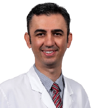
Education Information:
He graduated from Ankara University Faculty of Medicine in 2000 with the 4th rank. He completed his ophthalmology residency at Hacettepe University Faculty of Medicine . Between 2009 and 2011, he completed his master's degree in cornea, ocular surface and refractive surgery at Harvard Medical School in Boston, US. He qualified the title of associate professor in 2012 and Professor title in 2017 . He has performed over 15,000 surgeries in over twenty years.Academic achievements:
He has received numerous national and international awards (ARVO-Asia Travel Fellowship Grant Award, Gazi Eye Foundation 'Best Scientific Paper Award', ARVO Collaborative Research Fellowship Award, ICO Helmerich Fellowship Award, ICO WOC Fellowship Award, Royal College of) Ophthalmologists Best Reviewer Award, Turkish Academy of Sciences Outstanding Young Scientist Award). Koç University established its own research group with the support of KUTTAM, TÜBİTAK, TÜBA and ARVO.Awards, Memberships, Certificates:
- Ankara University Medical School, 4th rank in graduation (2000)
- International Council of Ophthalmology (ICO) “Basic Science Assessment in Ophthalmology including Optics and Refraction” Certificate (2004)
- International Council of Ophthalmology (ICO) “Clinical Science Assessment in Ophthalmology” Certificate (2005)
- Association for Research and Vision in Ophthalmology (ARVO)-Asia 'Travel Fellowship Grant Award' (2007)
- Gazi Eye Association Scientific Award (2008)
- TÜBİTAK ULAKBİM UBYT Awards
- Association for Research and Vision in Ophthalmology (ARVO) 'Collaborative Research Fellowship Award' (2009)
- TOD Glaucoma Symposium Best Oral Presentation Award (2009)
- TÜBİTAK 2219-Postdoctoral Research Fellow Award (2009)
- International Council of Ophthalmology (ICO) 'Helmerich International Fellowship Award' (2010)
- The Turkish Academy of Sciences (TÜBA) 'Outstanding Young Scientist Award' (2012)
- Association for Research and Vision in Ophthalmology (ARVO) 'Developing Country Eye Researcher Fellowship Award' (2015)
- Turkish Ophthalmology Society Scientific Award (2015)
- International Council of Ophthalmology (ICO) WOC2106 Travel Fellowship Grant Award (2016)
- Turkish Medical Association, Most Successful medical doctor of the year (2016)
- Turkish Ophthalmology Association
- Association for Research in Vision and Ophthalmology (ARVO)
- European Association for Vision and Eye Research (EVER)
- American Academy of Ophthalmology (AAO)
- American Society of Cataract and Refractive Surgery (ASCRS)
- European Society of Cataract and Refractive Surgeons (ESCRS)
Specialized Treatments and Surgeries:
- Corneal Transplant
- Complicated Cataract surgery
- Retinitis Pigmentosa Wharton jelly stem cell treatment
- Refractive Laser Eye Surgeries
- Keratoconus Cross-Linking
- Ring therapy
- Hard-hybrid innovative contact lenses
- Dry eye (Autologous serum, IPL, LipiFlow treatment)
- Glaucoma (eye pressure)
Foreign language:
- English
- Turkish
Harvard MEDICAL School
22 Years of EXPERIENCE
>15.000 Surgery
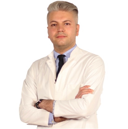
Education Information
He completed primary, secondary and high school in Baku. He graduated from Azerbaijan Medical Faculty in 2010. He completed his ophthalmology residency in Baku National Ophthalmology Center Azerbaijan . He was entitled to receive the Presidential Scholarship of the Republic of Azerbaijan in 2014, and completed the equivalence of specialization in Turkey in the Department of Ophthalmology, Faculty of Medicine, Selcuk University in 2014-2018, and worked as a research assistant and ophthalmologist at the university until 2018. He worked as an Ophthalmologist at Private LIV Hospital Nation between 2019-2022.Work experience:
- Selcuk University Faculty of Medicine
- Private LIV Hospital Ulus
Certificates, Memberships, Scientific Research:
- Peroperative developing choroidal detachment and its management.
- Surgical Approach in Posterior Polar Cataract.
- Iatrogenic retinal artery occlusion caused by cosmetic facial autologous fat filler injections.
- Effect of Smoking on Ocular Surface and Corneal Nerves.
- Lupus choroidopathy in a patient with discoid lupus erythematosus.
- Endophthalmitis and its treatment with early parsplanavitrectomy.
- Turkish Ophthalmology Association.
Specialized Treatments and Surgeries:
- Retractive Laser Eye Surgery: iLASIK (Femto Lasik), LASIK, LASEK, Trans Epithelial PRK (NO TOUCH) and SMILE
- Cataract Surgery (Smart Lens Surgery)
- Keratoconus Treatments – Cross-Linking Surgeries
- Pterygium Surgery
- Dry Eye Disease and Treatments
- Strabismus Surgery
- Glaucoma- Glaucoma Eye Pressure Treatments
- neuro ophthalmology
- Retinal Diseases Treatments
- Oculoplasty
- Uveitis Diseases
- Ectropion and entropion surgery – Eyelid deformity treatments
- Enucleation and Evisceration Prosthetic Eye Surgery
Foreign language:
- English
- German
- Russian
- Azerbaijani
- Turkish