Home »
Q-LASIK Laser Eye Surgery
Written and edited by the Smart Laser Eye Center Editorial Staff · Medically reviewed by Dr. Alim Huseynov, MD · Last updated: 10 September 2026
Q-LASIK is a flap-based laser vision correction procedure. It corrects myopia, hyperopia and astigmatism in the same way as conventional LASIK laser eye surgery, but the corneal flap is created with the Med-Logics ML7 microkeratome system, which was the first platform used to perform Q-LASIK. The excimer treatment that follows is identical in principle to any other LASIK procedure: the laser removes a calculated amount of corneal stroma so that light entering the eye focuses on the retina instead of in front of or behind it.
The letter Q refers to the quality control built into the flap step. Flap thickness, flap diameter, hinge position and vacuum level are not left to the moment of surgery; they are selected before the operation from the device nomogram, using the patient’s own steepest keratometry reading and horizontal corneal diameter.
What makes Q-LASIK different from standard LASIK
In every LASIK-family procedure the surgeon must first create a thin lamellar flap. How that flap is created is the single largest technical difference between the methods, and it is where Q-LASIK is distinct.
The ML7 is a mechanical microkeratome: a calibrated metal blade cuts a lamellar plane. A femtosecond laser, by contrast, does not cut. It places focused pulses that produce cavitation bubbles along a plane, and the flap is then dissected off that plane. The two approaches produce a flap by different physics, and they are described with different words throughout this page.
The following values are manufacturer specifications for the ML7 system, not results of an independent comparative trial:
- Flap creation in approximately 7 seconds, vacuum included. The complete cycle — suction ring application, vacuum, pass and release — is among the fastest in the LASIK field. A shorter suction time means a shorter period of raised intraocular pressure for the eye.
- Standard deviation of ±5 µm for the Med-Logics metal head used together with the Med-Logics CLB calibrated blade. A tight deviation matters because the residual stromal bed is calculated by subtracting the flap from the pachymetry: every micron of unexpected flap thickness is a micron taken away from the tissue that must remain.
- Flap thickness in 10 µm steps. With a 100 µm head, the calibrated blade series (−20, −10, plano, +10, +20) yields 80, 90, 100, 110 and 120 µm. The surgeon therefore selects the flap to fit the cornea, rather than fitting the cornea to a fixed flap.
- Hinge at any orientation between 0° and 180°. The suction ring is rotated by the surgeon, so the hinge can be placed temporally, nasally, superiorly, or on any oblique axis — including perpendicular to the astigmatic axis.
- Ring and vacuum from the device nomogram. Ring diameter follows the steepest K reading and the horizontal white-to-white measurement; vacuum follows the ring and the corneal thickness.
Why hinge direction is planned, not assumed
The corneal sub-basal nerve plexus is supplied mainly by fibres entering the cornea at the 3 and 9 o’clock positions. A temporal or nasal hinge preserves the largest proportion of these fibres during LASIK, because the flap edge runs across fewer of the entering trunks. A superior hinge crosses both of them.
Corneal sensation drives the tear reflex, so nerve preservation is directly linked to how dry the eye feels in the first months after surgery. In a randomised, self-controlled trial using mechanical microkeratomes, corneal sensation had returned to baseline by six months in eyes with a non-superior hinge, while eyes with a superior hinge had not recovered, and dry-eye signs and symptoms were more severe in that group.
Donnenfeld ED et al. 2003 · Ophthalmology 110(5):1023–9 · doi:10.1016/S0161-6420(03)00100-3. In series using femtosecond flaps this difference has not been reproduced — Mian SI et al. 2009 · J Cataract Refract Surg 35(12):2092–8 · doi:10.1016/j.jcrs.2009.07.009.
Temporal is the direction generally preferred at our centre. It preserves the nerve fibres described above and it also sits away from the frontal orbital rim, so the flap edge is less exposed in the direction from which blunt trauma most often arrives. A superior hinge may be chosen instead when the anatomy of the individual eye calls for it — for example a narrow palpebral aperture, a deep orbit, or a corneal curvature map in which the superior periphery gives the more favourable hinge geometry.
Hinge geometry is a calculation, not a preference. The cornea is not the same thickness under every meridian. Where the flap edge crosses a flatter zone, the flap thins. Our planning takes the steepest keratometry reading in the astigmatic axis and the lowest reading in the 8.5–9.0 mm ring under the intended hinge, converts the difference to microns (1 D of curvature corresponds to roughly 10 µm of thickness) and checks that what remains at the hinge is still robust. If it is not, the plan changes: the hinge moves to the steep axis, a thicker calibrated blade is selected, or the vacuum is adjusted — before the operation, not during it.
The numbers behind a Q-LASIK decision
Whether an eye can safely undergo Q-LASIK is not decided by the spectacle prescription. It is decided by how much corneal tissue the treatment consumes relative to how much that particular cornea has, and by what the corneal curvature will be once the treatment is finished. Four figures carry the decision.
- PTA — percent tissue altered. The flap plus the ablation depth, divided by the thinnest pachymetry. Our ceiling is 40 % when the steepest K is 46.00 D or above, and 45 % below that. If Kmax has not been measured, the stricter 40 % figure is applied.
- RSB — residual stromal bed. The stroma left underneath the flap after ablation. Floor for the LASIK family, Q-LASIK included: 300 µm.
- RCT — residual corneal thickness. Pachymetry minus ablation. Floor: 400 µm. RCT is independent of flap thickness, so a thinner flap improves RSB and PTA but never rescues RCT; only a shallower ablation does that.
- Post-operative keratometry window. The cornea must remain an effective lens after treatment: no flatter than 36 D and no steeper than 49 D. This is the limit that most often caps how much correction a flat cornea can carry, even when tissue is plentiful.
These thresholds are not arbitrary. In a case-control study of eyes that developed post-LASIK ectasia despite entirely normal pre-operative topography, 97 % had a PTA of 40 % or more, and PTA was the single most significant independent variable in multivariate analysis.
Santhiago MR et al. 2014 · Am J Ophthalmol 158(1):87–95 · doi:10.1016/j.ajo.2014.04.002
How RLES AI supports Q-LASIK planning
RLES AI® is refractive laser eye surgery planning software developed by our group. It is a clinical decision support tool: it reads the values printed on your topography, tomography and biometry reports, applies the rule set above, and returns the surgical options that pass every threshold together with the cost of each. It does not make the decision. The final clinical judgement always rests with your surgeon.
For a Q-LASIK plan the software works through, among others:
- Keratoconus and ectasia screening — BAD-D, ART max and average, Kmax, ISV and IVA, posterior elevation, pachymetric progression, and the biomechanical indices CBI and TBI when a Corvis ST measurement has been supplied. A borderline or pathological index does not simply raise a flag; it changes which procedures remain on the table. In an established or progressive ectatic cornea, corneal ablation is outside the rule set altogether — surface ablation included.
- Contact lens warpage detection — a set of indicators that separate a genuinely irregular cornea from one that has been temporarily deformed by lens wear. This distinction matters because a warped cornea can produce a false keratoconus pattern, and planning an ablation on a surface that will change shape once the lenses are stopped leads to residual refractive error.
- Tissue budgeting — PTA, RSB and RCT computed for every combination of flap thickness and optical zone that the device can deliver, with the combinations ranked by the residual stroma they leave.
- ML7 nomogram verification — suction ring, vacuum level and calibrated blade cross-checked against the steepest K and the corneal diameter, together with the hinge calculation described above for the temporal, superior and nasal directions separately.
- Maximum treatable correction — the dioptric ceiling imposed by the post-operative keratometry window for that specific cornea, which is frequently reached before the tissue thresholds are.
If the numbers do not permit Q-LASIK, the software says so and shows what does remain: TransEpithelial PRK, PRK, or a phakic ICL when the cornea is not the right place to carry the correction at all.
Read more about RLES AI® and its clinical rule set →
Who is a candidate for Q-LASIK
Q-LASIK may be considered when the patient is 18 years of age or older, the refraction has been stable, the cornea is topographically and tomographically normal, and the tissue thresholds above are met with margin. Between the ages of 18 and 21 the decision belongs to the surgeon, and documented refractive stability over at least twelve months is expected first.
Q-LASIK is not performed when there is keratoconus or suspicion of corneal ectasia, active ocular infection or inflammation, uncontrolled autoimmune or systemic disease, pregnancy or breastfeeding, severe untreated dry eye, or a cornea that cannot supply the required residual stroma. Several medications — isotretinoin and topiramate in particular — require the treatment to be postponed. Your medication history is asked for exactly for this reason.
Contact lens wear changes the shape of the cornea, and a warped cornea produces measurements that do not represent the eye you actually have. Soft lenses are usually stopped one to two weeks and rigid gas permeable lenses three to four weeks before measurement. The criterion, however, is not the calendar — it is two consecutive topographies that agree with each other.
Recovery after Q-LASIK
Because the epithelium is lifted with the flap rather than removed, the surface closes quickly and visual recovery is fast compared with surface ablation methods.
- First 5–6 hours. Mild to moderate stinging, watering and light sensitivity are usual. Resting with the eyes closed is the most useful thing a patient can do in this window.
- Day 1. Most patients see well enough for everyday tasks and attend the first post-operative examination.
- First week. No eye rubbing under any circumstances, and no water contact. Drops are used exactly as prescribed.
- Weeks 2–4. Swimming pools and the sea are avoided. Vision continues to sharpen and any fluctuation settles.
- Months 1, 3 and 6. Scheduled examinations. The one-month visit is the one that matters most for long-term corneal health.
Transient dry eye is the most common complaint after any flap-based procedure, because the flap interrupts corneal nerves. It typically improves over the first three to six months. Hinge planning, described earlier on this page, is one of the ways this effect is minimised.
Travelling to us for Q-LASIK
Patients who travel to Istanbul from another city or another country are usually able to complete the examination and the surgery within a short stay, provided that the pre-operative measurements are valid on arrival. Two conditions decide this: contact lenses must have been out for long enough for the cornea to have returned to its own shape, and any earlier reports must be sent in advance so that they can be reviewed before travel. The first post-operative examination is performed the day after surgery, and the following controls can be arranged with an ophthalmologist near the patient’s home, with the reports shared back to us.
Our patient concierge team arranges appointments, transfers and accommodation, and our patient journey page sets out what happens on each day.
Useful links
Frequently asked questions about Q-LASIK laser eye surgery
What is the difference between Q-LASIK and LASIK laser eye surgery?
The excimer treatment is the same. The difference is in how the corneal flap is created. Q-LASIK uses the Med-Logics ML7 microkeratome, which cuts the flap with a calibrated blade, completes the cycle including vacuum in about 7 seconds, and allows the hinge to be positioned at any orientation between 0° and 180°. Conventional LASIK may use another microkeratome or a femtosecond laser, which produces the plane by photodisruption so that the flap is then dissected rather than cut.
At what age can I have Q-LASIK laser eye surgery?
From 18 years of age. Between 18 and 21 the decision belongs to the surgeon, because refraction is less settled at that age; documented stability over at least twelve months is expected before proceeding. There is no fixed upper age limit, but from the mid-forties onwards presbyopia has to be discussed as part of the plan, and if a cataract has begun to form a lens-based procedure may be the more appropriate route.
Is Q-LASIK laser eye surgery painful?
The procedure itself is not painful. Anaesthetic drops are used; no injection and no general anaesthesia are involved. Pressure is felt during the few seconds of suction. For the first five to six hours afterwards, mild to moderate stinging, watering and light sensitivity are usual, and resting with the eyes closed during that period is the most helpful thing you can do. Flap-based procedures are markedly more comfortable in the first days than surface methods such as PRK, where the epithelium has to heal across the treated area.
How soon can I return to work after Q-LASIK laser eye surgery?
Most patients return to office work on the second or third day. Screen work is possible earlier than that but is tiring in the first days, and frequent breaks with lubricating drops help. Dusty environments, swimming pools and the sea are avoided for two to four weeks, and the eyes must not be rubbed at any point in the first week. Your surgeon sets the timetable for your own eyes at the day-one examination.
Can Q-LASIK be performed if I have keratoconus or a suspicious topography?
No. Established keratoconus and corneal ectasia are contraindications to corneal ablation, and this applies to surface methods as well, not only to flap procedures. A suspicious but not diagnostic topography is treated as a separate situation: further tests are requested, contact lens warpage is excluded, and the eye may be re-measured after an interval. If ectasia is confirmed, the discussion moves to corneal cross-linking and keratoconus treatment, and any refractive correction is considered only afterwards and by a different route.
What happens if my cornea is too thin for Q-LASIK laser eye surgery?
Thin does not automatically mean unsuitable; what matters is what remains after treatment. If the residual stromal bed or the residual corneal thickness would fall below the floors of 300 µm and 400 µm, or if the percent tissue altered would exceed the ceiling for your keratometry, then Q-LASIK is not planned for that eye. Alternatives are then assessed in order: a surface method such as TransEpithelial PRK, which does not consume tissue for a flap, or a phakic ICL, which carries the correction inside the eye and leaves the cornea untouched.
References
- Santhiago MR, Smadja D, Gomes BF, et al. Association between the percent tissue altered and post-laser in situ keratomileusis ectasia in eyes with normal preoperative topography. Am J Ophthalmol. 2014;158(1):87–95. doi:10.1016/j.ajo.2014.04.002
- Donnenfeld ED, Solomon K, Perry HD, et al. The effect of hinge position on corneal sensation and dry eye after LASIK. Ophthalmology. 2003;110(5):1023–9. doi:10.1016/S0161-6420(03)00100-3
- Mian SI, Li AY, Dutta S, Musch DC, Shtein RM. Dry eyes and corneal sensation after laser in situ keratomileusis with femtosecond laser flap creation: effect of hinge position, hinge angle, and flap thickness. J Cataract Refract Surg. 2009;35(12):2092–8. doi:10.1016/j.jcrs.2009.07.009
- Talamo JH, Meltzer J, Gardner J. Reproducibility of flap thickness with IntraLase FS and Moria LSK-1 and M2 microkeratomes. J Refract Surg. 2006;22(6):556–61. doi:10.3928/1081-597X-20060601-07
Device figures given on this page for the Med-Logics ML7 system are manufacturer specifications and are identified as such in the text. This page is patient information and does not replace an examination. Whether a procedure is suitable for a particular eye can only be established after a complete ophthalmological assessment, and the final clinical decision rests with your surgeon.
This page is general information only and is not a diagnosis or a treatment recommendation; suitability is determined by an eye examination.
Your Expert Eye Surgeons
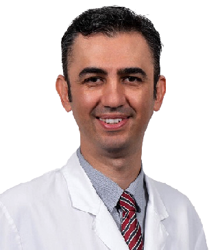
Education Information:
He graduated from Ankara University Faculty of Medicine in 2000 with the 4th rank. He completed his ophthalmology residency at Hacettepe University Faculty of Medicine . Between 2009 and 2011, he completed his master's degree in cornea, ocular surface and refractive surgery at Harvard Medical School in Boston, US. He qualified the title of associate professor in 2012 and Professor title in 2017 . He has performed over 15,000 surgeries in over twenty years.Academic achievements:
He has received numerous national and international awards (ARVO-Asia Travel Fellowship Grant Award, Gazi Eye Foundation 'Best Scientific Paper Award', ARVO Collaborative Research Fellowship Award, ICO Helmerich Fellowship Award, ICO WOC Fellowship Award, Royal College of) Ophthalmologists Best Reviewer Award, Turkish Academy of Sciences Outstanding Young Scientist Award). Koç University established its own research group with the support of KUTTAM, TÜBİTAK, TÜBA and ARVO.Awards, Memberships, Certificates:
- Ankara University Medical School, 4th rank in graduation (2000)
- International Council of Ophthalmology (ICO) “Basic Science Assessment in Ophthalmology including Optics and Refraction” Certificate (2004)
- International Council of Ophthalmology (ICO) “Clinical Science Assessment in Ophthalmology” Certificate (2005)
- Association for Research and Vision in Ophthalmology (ARVO)-Asia 'Travel Fellowship Grant Award' (2007)
- Gazi Eye Association Scientific Award (2008)
- TÜBİTAK ULAKBİM UBYT Awards
- Association for Research and Vision in Ophthalmology (ARVO) 'Collaborative Research Fellowship Award' (2009)
- TOD Glaucoma Symposium Best Oral Presentation Award (2009)
- TÜBİTAK 2219-Postdoctoral Research Fellow Award (2009)
- International Council of Ophthalmology (ICO) 'Helmerich International Fellowship Award' (2010)
- The Turkish Academy of Sciences (TÜBA) 'Outstanding Young Scientist Award' (2012)
- Association for Research and Vision in Ophthalmology (ARVO) 'Developing Country Eye Researcher Fellowship Award' (2015)
- Turkish Ophthalmology Society Scientific Award (2015)
- International Council of Ophthalmology (ICO) WOC2106 Travel Fellowship Grant Award (2016)
- Turkish Medical Association, Most Successful medical doctor of the year (2016)
- Turkish Ophthalmology Association
- Association for Research in Vision and Ophthalmology (ARVO)
- European Association for Vision and Eye Research (EVER)
- American Academy of Ophthalmology (AAO)
- American Society of Cataract and Refractive Surgery (ASCRS)
- European Society of Cataract and Refractive Surgeons (ESCRS)
Specialized Treatments and Surgeries:
- Corneal Transplant
- Complicated Cataract surgery
- Retinitis Pigmentosa Wharton jelly stem cell treatment
- Refractive Laser Eye Surgeries
- Keratoconus Cross-Linking
- Ring therapy
- Hard-hybrid innovative contact lenses
- Dry eye (Autologous serum, IPL, LipiFlow treatment)
- Glaucoma (eye pressure)
Foreign language:
- English
- Turkish
Harvard MEDICAL School
22 Years of EXPERIENCE
>15.000 Surgery
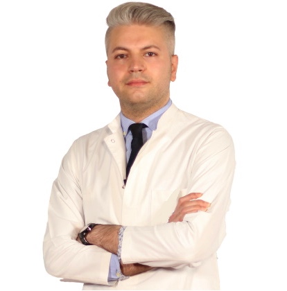
Education Information
He completed primary, secondary and high school in Baku. He graduated from Azerbaijan Medical Faculty in 2010. He completed his ophthalmology residency in Baku National Ophthalmology Center Azerbaijan . He was entitled to receive the Presidential Scholarship of the Republic of Azerbaijan in 2014, and completed the equivalence of specialization in Turkey in the Department of Ophthalmology, Faculty of Medicine, Selcuk University in 2014-2018, and worked as a research assistant and ophthalmologist at the university until 2018. He worked as an Ophthalmologist at Private LIV Hospital Nation between 2019-2022.Work experience:
- Selcuk University Faculty of Medicine
- Private LIV Hospital Ulus
Certificates, Memberships, Scientific Research:
- Peroperative developing choroidal detachment and its management.
- Surgical Approach in Posterior Polar Cataract.
- Iatrogenic retinal artery occlusion caused by cosmetic facial autologous fat filler injections.
- Effect of Smoking on Ocular Surface and Corneal Nerves.
- Lupus choroidopathy in a patient with discoid lupus erythematosus.
- Endophthalmitis and its treatment with early parsplanavitrectomy.
- Turkish Ophthalmology Association.
Specialized Treatments and Surgeries:
- Retractive Laser Eye Surgery: iLASIK (Femto Lasik), LASIK, LASEK, Trans Epithelial PRK (NO TOUCH) and SMILE
- Cataract Surgery (Smart Lens Surgery)
- Keratoconus Treatments – Cross-Linking Surgeries
- Pterygium Surgery
- Dry Eye Disease and Treatments
- Strabismus Surgery
- Glaucoma- Glaucoma Eye Pressure Treatments
- neuro ophthalmology
- Retinal Diseases Treatments
- Oculoplasty
- Uveitis Diseases
- Ectropion and entropion surgery – Eyelid deformity treatments
- Enucleation and Evisceration Prosthetic Eye Surgery
Foreign language:
- English
- German
- Russian
- Azerbaijani
- Turkish