Home »
Keratoconus Cross-Linking Surgery
Keratoconus Disease – Cross-Linking Surgery
Written and edited by the Smart Laser Eye Center Editorial Staff · Medically reviewed by Dr. Alim Huseynov, MD and Prof. Dr. Levent Alimgil, MD · Last updated: 10 September 2026
Keratoconus Cross-Linking
Especially, Keratoconus Disease causes blurry vision of the cornea, which develops in the shape of an outward cone, and may cause sensitivity to light and glare. Usually affects both eyes, but often affects one eye more than the other. Also, it usually begins to affect people between the ages of 10 and 25. Additionally, the condition may progress slowly for 10 years or more. However, the development of keratoconus usually slows down and stops after the age of 35. Also, Keratoconus Cross-Linking Surgery very effective treatment of Keratoconus disease.
In the early stages of keratoconus, you can correct vision problems with glasses or soft contact lenses. You may then need to continue treatment with other types of lenses, such as rigid, permeable contact lenses or scleral lenses. If there is no treatment and your condition progresses to an advanced stage, you may need a corneal transplant.
Treatment Method
Cross-linking surgery is performing to increase the biomechanical stability and stiffness of the cornea in order to prevent the progression of keratoconus. Technically, in the center of the eye, with a diameter of approximately 6-7 mm, the epithelium, which is the top layer of the cornea, is strip and 0.1% Riboflavin solution (also known as vitamin B₂ as Riboflavin content) to be drip into the eye, ultraviolet-A radiation light at a wavelength of 370 nm is apply in accordance with a certain procedure.
It is done with the application by the doctor. Ultraviolet-A radiation light activates Riboflavin, leading to the formation of collagen bonds between collagen fibrils and corneal stroma and an intense healing process in the anterior stroma. More importantly, This technique should not be apply to patients with a central corneal thickness of less than 400 microns, as it may cause a risk of toxic reactions in the endothelium, the innermost layer of the cornea.
Laser Eye Treatment
According to the condition of the cornea and eye structure, in patients with keratoconus whose progression has stopped after the age of 35, first Smart Laser Eye surgery and immediately after cross-linking surgery can be perform and the patient can get rid of his glasses or lenses. Keratoconus Patients are treat under the Sensitive Profile Patient Program.
ICL (Intraocular) Phakic lenses can be a very effective solution in the treatment of eye defects in patients who have had Cross-Linking treatment in Keratoconus Disease and whose development of Keratoconus disease has stopped. Click for information about ICL (Intraocular Lens)…
These treatments can be offer in addition to the above vision correction options, depending on the patient’s condition.
Symptoms
Although keratoconus is of varying severity, it typically affects both eyes and has well-known signs and symptoms, although there is no clear consensus on the signs and symptoms associated with early keratoconus. And these findings can be detect with a state-of-the-art corneal tomography device. The early stages of the disease are often referred to as subclinical or form-fruste keratoconus, but there are no unified criteria for the use of these two terms.
Subclinical keratoconus is typically a reference to one eye with normal corneal hourglass findings and topographic signs of keratoconus (or questionable topographic findings) in the other eye with keratoconus.
The signs and symptoms of keratoconus may change as the disease progresses.
- Blurred or distorted vision.
- Increased sensitivity to bright light and glare, which can cause problems when driving at night.
- The need for frequent changes in eyeglass prescriptions.
- Sudden worsening or blurred vision.
When to see a doctor
If your eyesight is deteriorating rapidly due to astigmatism, you should definitely call our hospital and see your ophthalmologist. During routine eye examinations in our hospital, especially keratoconus symptoms are analyze and keratoconus risk is determined.
Causes of the Disease
Although the cause cannot be determine exactly, among the causes of the disease in medical research; It has been determined that genetic and environmental factors from the family are caused by asthma, eczema, allergies, intense eye rubbing. Keratoconus most often appears with no family history at all, but a minority of patients do have an affected relative, and having a close relative with keratoconus raises your own risk — which is why it is worth screening siblings and children.
Lucas SEM, Burdon KP. Genetic and Environmental Risk Factors for Keratoconus. Annu Rev Vis Sci. 2020;6:25-46. doi:10.1146/annurev-vision-121219-081723
Risk factors
These factors can increase your chances of developing keratoconus:
- Having a family history of keratoconus
- rub your eyes vigorously
- Having certain conditions such as retinitis pigmentosa, Down syndrome, Ehlers-Danlos syndrome, hay fever, and asthma
Complications
In some cases, your cornea can swell rapidly, causing a sudden decrease in vision and scarring of the cornea. This is cause by a condition where the inner lining of your cornea breaks down, allowing fluid to enter the cornea (hydrops). The swelling usually goes away on its own, but a scar may form that affects your vision.
Advanced keratoconus can cause injury to your cornea, especially where the cone is most prominent. An injured cornea causes worsening vision problems and may require corneal transplant surgery.
Diagnosis
To diagnose keratoconus, our ophthalmologist will review your medical and family history and perform an eye exam. He or she may do other tests to determine more details about the shape of your cornea. Tests to diagnose keratoconus include:
Eye breakout. In this test, your eye doctor uses special equipment that measures your eyes to check for vision problems. He may ask you to look at a device with different lenses to help you determine which combination gives you the sharpest vision. He or she may also use a retinoscope to evaluate your eyes.
Examination with a bio-microscope (Slit-Lamp). In this test, your doctor directs a vertical beam of light to the surface of your eye and uses a low-power microscope to view your eye. It evaluates the shape of your cornea and looks for other potential problems with your eye.
Keratometry Test
In this test, your eye doctor focuses a circle of light on your cornea and measures the reflection to determine the basic shape of your cornea.
Computerized corneal mapping
Special photographic tests such as corneal tomography and corneal topography record images to create a detailed shape map of your cornea. A corneal CT can also measure the thickness of your cornea. Corneal tomography can detect early signs of keratoconus before the disease appears during examination with the Bio-Microscope.
Treatment options in keratoconus: cross-linking, CAIRS and ring segments
Keratoconus treatment answers two separate questions, and the two should not be confused. The first is whether the disease can be stopped from progressing; the second is whether the quality of vision can be improved. Corneal cross-linking answers the first question, and intrastromal ring procedures answer the second. For that reason the two are usually not alternatives to one another but consecutive steps of the same plan.
The first question: is the disease progressing?
Progression is not established from a single measurement. According to the current international consensus, at least two of the following three findings must be documented together: steepening of the anterior corneal surface (an increase of 1.0 D or more in Kmax within 12 months), steepening of the posterior surface, and thinning of the cornea. A single topography taken at the first visit is therefore rarely sufficient; consecutive measurements on the same device are required.
Below the age of 18 the situation is different. Because the disease can progress considerably faster in children and adolescents, cross-linking is not delayed in this group. Conversely, prophylactic cross-linking of an adult eye that shows no documented progression is not supported by current evidence.
Gomes JAP, Tan D, Rapuano CJ, et al. Global consensus on keratoconus and ectatic diseases. Cornea. 2015;34(4):359-69. doi:10.1097/ICO.0000000000000408
Cross-linking: which protocol, and at which corneal thickness?
Corneal cross-linking stiffens the cornea by combining riboflavin with ultraviolet-A light. Its purpose is not to correct vision but to arrest progression. In the original Dresden series, 23 eyes with progressive keratoconus were treated at 3 mW/cm² for 30 minutes; progression was halted in every eye, and in 70 per cent of them maximum keratometry regressed by a mean of 2.01 D. Which protocol may be applied is decided by corneal thickness:
- 400 µm and above — the conventional (Dresden) protocol at 3 mW/cm² for 30 minutes, or an accelerated protocol at 9 mW/cm² for 10 minutes. Both deliver the same total dose of 5.4 J/cm².
- 320–399 µm — thickness is temporarily raised with hypo-osmolar riboflavin or with a contact lens-assisted technique, and 400 µm is confirmed intraoperatively before the ultraviolet phase is started.
- Below 320 µm — cross-linking is not performed. Other options are evaluated in these eyes.
This limit is not arbitrary. In a 400 µm cornea saturated with riboflavin the irradiance reaching the inner cell layer of the cornea is approximately 0.18 mW/cm², which is about half of the 0.35 mW/cm² damage threshold measured for endothelial cells. Cross-linking depopulates the stroma of keratocytes to a depth of roughly 300 µm, and that zone repopulates over a period of up to six months. As the cornea thins, the safety margin at the endothelium disappears first, which is precisely why the choice of protocol is tied to thickness.
Wollensak G, Spoerl E, Seiler T. Am J Ophthalmol. 2003;135(5):620-7. doi:10.1016/s0002-9394(02)02220-1 · Spoerl E, Mrochen M, Sliney D, Trokel S, Seiler T. Cornea. 2007;26(4):385-9. doi:10.1097/ICO.0b013e3180334f78
An accelerated protocol is not simply the same treatment delivered faster. In a meta-analysis of 14 randomised controlled trials, the conventional protocol produced greater corneal flattening and a deeper stromal demarcation line, whereas the accelerated protocol caused less reduction in central corneal thickness. No difference was found between the two in uncorrected or corrected visual acuity, spherical equivalent, corneal biomechanical measurements or endothelial cell density. In other words the two protocols are close but not interchangeable, and the choice belongs to the surgeon.
Yeh CY, Chen KC, Chen YJ, Cheng SF. Eur J Ophthalmol. 2025;35(3):785-798. doi:10.1177/11206721241298317
For thin corneas the contact lens-assisted technique has been described in measured terms: in a series of 14 eyes whose minimum thickness after epithelial removal was 377.2 ± 14.5 µm, a riboflavin-soaked soft contact lens raised the functional thickness to 485.1 ± 15.8 µm, a mean gain of 107.9 µm. The stromal demarcation line was measured at 252.9 ± 40.8 µm and no significant endothelial cell loss was recorded.
Jacob S, Kumar DA, Agarwal A, et al. J Refract Surg. 2014;30(6):366-72. doi:10.3928/1081597X-20140523-01
CAIRS: a segment prepared from donor cornea instead of synthetic material
CAIRS stands for Corneal Allogenic Intrastromal Ring Segments. The distinguishing feature of the method is that the segment placed inside the cornea is not synthetic: it is trephined from donated human corneal tissue. The segment is implanted into a channel dissected within the corneal stroma, and by flattening the cone it aims to improve the quality of vision rather than to stop the disease.
Compared with synthetic rings, the most consistently reported difference is that extrusion of the implant and melting of the tissue around it occur markedly less often, because the implanted material has the same structure as the cornea itself. CAIRS has also been applied in thinner corneas than synthetic segments allow; published series report a lower bound in the region of 320–350 µm. Channel depth is generally taken as 35–70 per cent of corneal thickness or as a fixed 200–250 µm, with a segment width of about 1.5 mm. There is no universal nomogram: several groups have published their own.
In the series that introduced the technique, 24 eyes with stage 1 to 4 keratoconus received segments in the 6.5 mm optic zone through femtosecond-dissected channels, followed by accelerated cross-linking, or by the contact lens-assisted variant where the cornea was too thin. Over a mean follow-up of 11.58 ± 3.6 months uncorrected distance visual acuity improved by 2.79 ± 2.65 lines and corrected distance visual acuity by 1.29 ± 1.33 lines; no eye progressed and no segment-induced complication was observed. This was a pilot study, and its authors state plainly that long-term outcomes remain to be determined.
Jacob S, Patel SR, Agarwal A, et al. J Refract Surg. 2018;34(5):296-303. doi:10.3928/1081597X-20180223-01
ICRS: synthetic intrastromal corneal ring segments
ICRS are synthetic segments implanted inside the cornea. They flatten the cone, reduce irregular astigmatism and in most cases improve what the eye can achieve with spectacles or a contact lens. The thickness of the segment, the arc length and the position of implantation are selected according to corneal topography, the location of the cone and the astigmatism to be corrected.
One point deserves emphasis here: planning depends on the brand of segment used. Every manufacturer publishes its own nomogram and its own optic zone — some segments are designed for a 5.0 to 6.0 mm optic zone, others for a fixed 5.0 mm, and others again for diameters of 6.7 to 8.2 mm. A segment plan therefore cannot be produced before the brand has been decided. Channel depth is typically dissected at 75 to 80 per cent of corneal thickness, and the entry incision is placed on the steep meridian.
Vega-Estrada A, Alio JL. Eye Vis (Lond). 2016;3:8. doi:10.1186/s40662-016-0040-z
Sequencing: which treatment comes first?
- In progressive keratoconus the ring procedure and cross-linking are most often performed in the same session.
- In a cornea that has already been cross-linked, the flattening effect of a segment is reduced, because the tissue has been stiffened. The sequence therefore has to be planned from the outset rather than decided later.
- Where Kmax exceeds 70 D, or where there is scarring in the centre of the cornea, the contribution of segments and cross-linking is limited; in these eyes corneal transplantation (DALK or penetrating keratoplasty) comes into consideration.
- At least 6 to 12 months of documented stability is expected before the residual refraction is addressed. In suitable eyes an intraocular lens (phakic ICL) may then be evaluated.
Which of these options is appropriate for your eye is decided by your surgeon, after all of the measurements have been evaluated together.
Standard refractive laser treatment is not performed on an eye with established or progressing keratoconus. This applies to surface ablation as well: the restriction is not a property of the flap but of the tissue that is removed. Cross-linking, ring segments and, where required, transplantation address the disease itself; refractive correction is discussed only after stability has been documented.
How many items does one keratoconus assessment require?
In keratoconus the decision does not rest on a single map. It rests on a data set whose items check one another. For two eyes, the items that have to be collected, read and compared are distributed as follows:
| History and questionnaire items | 16 / 30 |
| Examination findings | 27 / 50 |
| Values read from device printouts | 84 |
| Calculations performed | 140 |
| Thresholds and nomogram rows to be recalled | ≈130 |
| Total items checked in every case | ≈300 |
Most of these items are ones the surgeon already knows. The difficult part is not knowing them; it is interrogating every one of them, in the same order, without exception, in every case. At the end of a busy day, a single line skipped is a far more realistic risk than a gap in knowledge.
What RLES AI screens in this plan
Our clinic uses RLES AI®, a clinical decision support system developed for refractive and corneal surgical planning, as a second pass over the keratoconus assessment. It does not replace the examination; it makes sure that what is knowable is not overlooked.
- Keratoconus and ectasia screening — tomographic indices are read together. Indices that are mathematically dependent on one another are not counted as separate pieces of evidence, which reduces both false alarms and false reassurance.
- Contact lens-induced surface change — lens wear can temporarily distort corneal shape. Where that pattern is present the measurements are treated as unreliable and the plan is not finalised until consecutive, mutually consistent maps are obtained.
- Ocular surface and dry eye — untreated dry eye both distorts the measurement and increases symptoms, so the surface is stabilised first.
- Intraocular pressure and anterior chamber angle — the angle value on the preoperative tomography is read in every case, and a narrow angle is referred for separate assessment.
Beyond screening, the software checks whether the intended plan is compatible with the measurements of that particular cornea:
- Tissue safety engine — the thickness that will remain after treatment is calculated and compared with the safety floors.
- Keratoconus-specific checks — the progression criteria (anterior and posterior steepening, thinning) are compared across consecutive measurements; for cross-linking the thinnest corneal thickness is checked against the protocol thresholds above, and a ring plan is verified against the nomogram of the specific segment selected.
- Preconditions for complications — medication history and chronic disease questions are worked through in the same order in every case.
What this screening gives you
The benefit is this: the decision is made by your surgeon, but it is on record that every one of these items was asked before the decision was taken. If an item was not asked or not measured, the report names it as not reported rather than leaving it blank. No missing item is quietly counted as normal.
The software does not make a diagnosis, does not decide on surgery and does not guarantee an outcome. Detailed information about the system is available at rlesai.com.
Useful links:
About our eye surgeons. Presbyopia LASIK Laser Eye Surgery. LASIK Laser Eye Surgery. NoTouch T-PRK Laser Eye Surgery. PRK Laser Eye Surgery. Sensitive Profile Patient Program. LASIK Definition. FAQ.
Frequently Ask Questions about Keratoconus Treatment
At what age can I have Cross-Linking Surgery?
Keratoconus patients can have Cross-Linking Eye surgeries at any age, regardless of their age, to stop or slow the progression of the disease, according to your Doctor’s decision.
I have keratoconus, should I have Cross-Linking Surgery?
If you are a keratoconus patient and want to stop the progression of the disease, you should have Cross-Linking Surgery according to your doctor’s advice. In this way, the progress of your eye numbers will be stopped and slowed down.
I have keratoconus. Can I have Laser Eye Surgery?
Laser Eye Surgery
If you are a Keratoconus patient and you have reached the age of 35, it is expected that your Keratoconus development will stop or slow down. If your doctor determines that your Keratoconus disease has stopped and your eye is medically suitable for Smart Laser Eye Surgery, you can get rid of your glasses as Laser Eye Surgery. Having Cross-linking surgery before the age of 35 will give you an advantage. Because the lower your number, the higher your chance of meeting the corneal conditions for Laser Surgery. Especially in the case of Keratoconus patients who have laser eye surgery after the age of 35, Cross-linking surgery should be performed immediately after the Laser Eye Surgery to strengthen the corneal ligaments one more time for the future.
How much does Cross-Linking Surgery cost?
Keratoconus Cross-Linking cost per eye is applied in Cross-Linking surgeries. For more pricing information please click the link..
This page is general information only and is not a diagnosis or a treatment recommendation; suitability is determined by an eye examination.
Your Expert Eye Surgeons
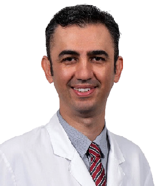
Education Information:
He graduated from Ankara University Faculty of Medicine in 2000 with the 4th rank. He completed his ophthalmology residency at Hacettepe University Faculty of Medicine . Between 2009 and 2011, he completed his master's degree in cornea, ocular surface and refractive surgery at Harvard Medical School in Boston, US. He qualified the title of associate professor in 2012 and Professor title in 2017 . He has performed over 15,000 surgeries in over twenty years.Academic achievements:
He has received numerous national and international awards (ARVO-Asia Travel Fellowship Grant Award, Gazi Eye Foundation 'Best Scientific Paper Award', ARVO Collaborative Research Fellowship Award, ICO Helmerich Fellowship Award, ICO WOC Fellowship Award, Royal College of) Ophthalmologists Best Reviewer Award, Turkish Academy of Sciences Outstanding Young Scientist Award). Koç University established its own research group with the support of KUTTAM, TÜBİTAK, TÜBA and ARVO.Awards, Memberships, Certificates:
- Ankara University Medical School, 4th rank in graduation (2000)
- International Council of Ophthalmology (ICO) “Basic Science Assessment in Ophthalmology including Optics and Refraction” Certificate (2004)
- International Council of Ophthalmology (ICO) “Clinical Science Assessment in Ophthalmology” Certificate (2005)
- Association for Research and Vision in Ophthalmology (ARVO)-Asia 'Travel Fellowship Grant Award' (2007)
- Gazi Eye Association Scientific Award (2008)
- TÜBİTAK ULAKBİM UBYT Awards
- Association for Research and Vision in Ophthalmology (ARVO) 'Collaborative Research Fellowship Award' (2009)
- TOD Glaucoma Symposium Best Oral Presentation Award (2009)
- TÜBİTAK 2219-Postdoctoral Research Fellow Award (2009)
- International Council of Ophthalmology (ICO) 'Helmerich International Fellowship Award' (2010)
- The Turkish Academy of Sciences (TÜBA) 'Outstanding Young Scientist Award' (2012)
- Association for Research and Vision in Ophthalmology (ARVO) 'Developing Country Eye Researcher Fellowship Award' (2015)
- Turkish Ophthalmology Society Scientific Award (2015)
- International Council of Ophthalmology (ICO) WOC2106 Travel Fellowship Grant Award (2016)
- Turkish Medical Association, Most Successful medical doctor of the year (2016)
- Turkish Ophthalmology Association
- Association for Research in Vision and Ophthalmology (ARVO)
- European Association for Vision and Eye Research (EVER)
- American Academy of Ophthalmology (AAO)
- American Society of Cataract and Refractive Surgery (ASCRS)
- European Society of Cataract and Refractive Surgeons (ESCRS)
Specialized Treatments and Surgeries:
- Corneal Transplant
- Complicated Cataract surgery
- Retinitis Pigmentosa Wharton jelly stem cell treatment
- Refractive Laser Eye Surgeries
- Keratoconus Cross-Linking
- Ring therapy
- Hard-hybrid innovative contact lenses
- Dry eye (Autologous serum, IPL, LipiFlow treatment)
- Glaucoma (eye pressure)
Foreign language:
- English
- Turkish
Harvard MEDICAL School
22 Years of EXPERIENCE
>15.000 Surgery
Education Information
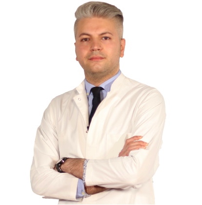
He completed primary, secondary and high school in Baku. He graduated from Azerbaijan Medical Faculty in 2010. He completed his ophthalmology residency in Baku National Ophthalmology Center Azerbaijan . He was entitled to receive the Presidential Scholarship of the Republic of Azerbaijan in 2014, and completed the equivalence of specialization in Turkey in the Department of Ophthalmology, Faculty of Medicine, Selcuk University in 2014-2018, and worked as a research assistant and ophthalmologist at the university until 2018. He worked as an Ophthalmologist at Private LIV Hospital Nation between 2019-2022.Work experience:
- Selcuk University Faculty of Medicine
- Private LIV Hospital Ulus
Certificates, Memberships, Scientific Research:
- Peroperative developing choroidal detachment and its management.
- Surgical Approach in Posterior Polar Cataract.
- Iatrogenic retinal artery occlusion caused by cosmetic facial autologous fat filler injections.
- Effect of Smoking on Ocular Surface and Corneal Nerves.
- Lupus choroidopathy in a patient with discoid lupus erythematosus.
- Endophthalmitis and its treatment with early parsplanavitrectomy.
- Turkish Ophthalmology Association.
Specialized Treatments and Surgeries:
- Retractive Laser Eye Surgery: iLASIK (Femto Lasik), LASIK, LASEK, Trans Epithelial PRK (NO TOUCH) and SMILE
- Cataract Surgery (Smart Lens Surgery)
- Keratoconus Treatments – Cross-Linking Surgeries
- Pterygium Surgery
- Dry Eye Disease and Treatments
- Strabismus Surgery
- Glaucoma- Glaucoma Eye Pressure Treatments
- neuro ophthalmology
- Retinal Diseases Treatments
- Oculoplasty
- Uveitis Diseases
- Ectropion and entropion surgery – Eyelid deformity treatments
- Enucleation and Evisceration Prosthetic Eye Surgery
Foreign language:
- English
- German
- Russian
- Azerbaijani
- Turkish
MEDICAL UNIVERSITY of Selcuk
12 Years of EXPERIENCE
>8.000 Surgery
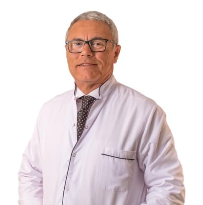
Prof. Dr. Levent Alimgil graduated from Istanbul University Cerrahpasa Faculty of Medicine in 1986 and completed his residency in ophthalmology at Trakya University Faculty of Medicine in 1990. He was appointed Assistant Professor in 1994, Associate Professor in 2001 and Professor of Ophthalmology in 2007 at the same faculty.
He served as Medical Director of the Yeditepe University Eye Research Centre between 2007 and 2009, and as Head of the Department of Ophthalmology at Medicana International Istanbul Hospital between 2009 and 2019. Since 2025 he has been Medical Director of the Nisantasi Hospital Eye Centre.
His clinical practice covers cataract surgery and refractive lens exchange with monofocal, multifocal, trifocal and toric intraocular lenses, phakic ICL implantation, keratoconus management and corneal cross-linking, the medical and surgical treatment of glaucoma including trabeculectomy, and corneal transplantation. He has performed more than 6,000 keratoplasty procedures.
He is a member of the Turkish Ophthalmological Association and of the American Academy of Ophthalmology (AAO). He speaks English and German.
ISTANBUL UNIVERSITY Cerrahpasa
36 Years of EXPERIENCE
>6.000 Corneal Transplants
Patients Reviews
Definitely avoid low-cost Cross-Linking Surgery
You may think that a cheap Cross-Linking surgery is right for you. This might be fine if you're buying a cheap TV, but it's not worth the gamble with your eyesight. But as you know, having cheap eye surgery means sacrificing technology, physician quality, medical care and sterile conditions, and most importantly, taking risks. The issues that fall on a patient who wants to have cataract surgery and should pay the most attention; The hospital with the latest technology in cataract surgery and imaging devices, a sterile environment, and an experienced doctor and clinical team should be selected. We would like to remind all our patients that they only have two eyes and that the most important and most sensitive sense organ is their eyes.
None of these things are more important than your eye health, and we do not compromise on quality and cutting-edge technology. We offer you our prices in a very understandable, fair and affordable way.
There are no hidden costs in our pricing. We make Cross-Linking Surgery affordable for you.